Project: Modeling and visualizing diffusion MRI data
Description

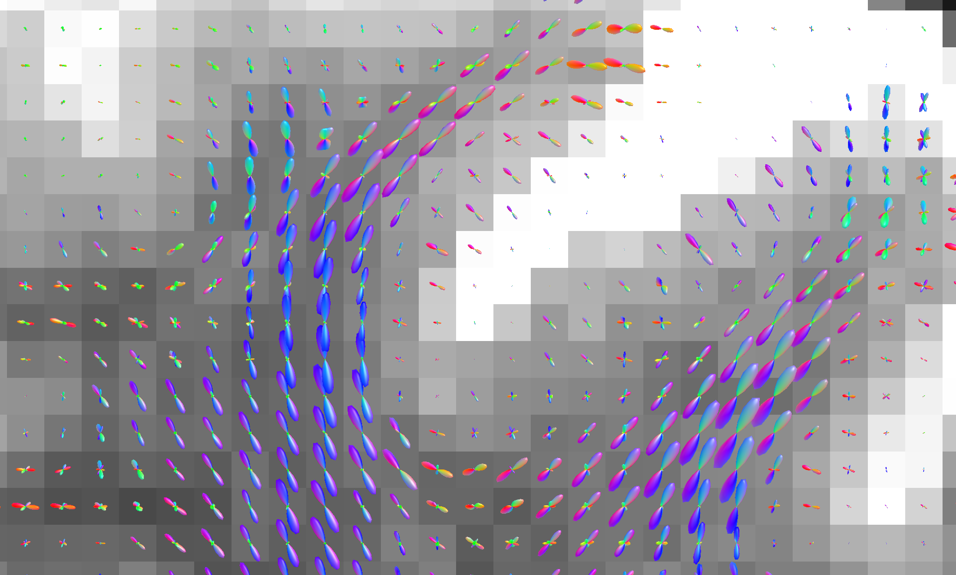
Diffusion MRI allows us to make spectacular visualizations of the human brain. Depending on how you model the data coming out of the MRI scanner, you can obtain insights into different properties of the underlying tissue. Modeling fiber orientation distributions allows us to perform accurate fiber tracking and create the colorful, spaghetti-like images you might have seen before. Meanwhile, a more complex microstructural model, such as the 'Standard Model,' can provide information about physiological changes at a potentially microscopic scale.
Currently, most models are fit on a voxel level because the measurements are performed at this scale. Voxel-wise fitting has two issues: it disregards the inter-voxel correlation and often requires you to interpolate between voxels to get necessary values for downstream tasks.
We use implicit neural representations, which you might have seen in 3D rendering (NeRFs), data compression, or any other fields in which they are used. We use a spatial encoding and a simple neural network to represent the dataset. Doing this allows us to make use of the spatial correlations in the dataset and model the dataset as a continuous space.
So far, we have presented a basic version of our work at a conference and a more elaborate paper under review. There is still a lot of work to do, though. If you are excited by the prospect of using machine learning to model datasets, are interested in the brain, and want to help us explore the possibilities of this new direction in diffusion MRI modeling, contact us, and we can chat!
[1] Hendriks, T., Vilanova, A., & Chamberland, M. (2023, October). Neural Spherical Harmonics for structurally coherent continuous representation of diffusion MRI signal. In International Workshop on Computational Diffusion MRI (pp. 1-12). Cham: Springer Nature Switzerland.
[2] Hendriks, T., Vilanova, A., & Chamberland, M. (2024). Implicit Neural Representation of Multi-shell Constrained Spherical Deconvolution for Continuous Modeling of Diffusion MRI. bioRxiv, 2024-08.
Details
- Student
-
HPHoria Pivniceru
- Supervisor
-
 Maxime Chamberland
- Secondary supervisor
-
 Tom Hendriks